Media Center
A multimedia mosaic of moments at GIST
GIST Excellence
[Press release] Professor Sung Chan Jun"s research team demonstrates the "quasi-invasive brain electrical stimulation" effect with computermodeling
- 엘리스 리
- REG_DATE : 2017.02.01
- HIT : 1102
Professor Sung Chan Jun"s research team demonstrates the
"quasi-invasive brain electrical stimulation" effect with computer modeling

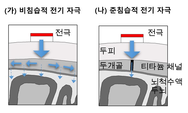
[Fig. 1] (a) In the case of non-invasive stimulation, current is spread by the scalp, skull, cerebrospinal fluid, etc., so that only a small amount of current reaches the brain. On the other hand, in the case of quasi-invasive electric stimulation, the electric current is transmitted into the brain via the titanium channel, so that a high electrical stimulus can be applied to the desired brain region.
□ Professor Sung Chan Jun of GIST (School of Electrical Engineering and Computer Science) has succeeded in demonstrating the effectiveness of "semi-invasive electric stimulation," which is attracting attention as a new brain disease treatment.
Using a computer simulation technique developed by the team, the researchers demonstrated that "quasi-invasive electrical stimulation" has a stimulation efficiency that is 10 times higher than "non-invasive brain stimulation," which transmits electric stimulation near the scalp or head.
This achievement is expected to contribute to the clinical application of "quasi-invasive electrical stimulation" that can treat brain diseases by safe and efficient electrical stimulation of the brain.
□ "Brain electrical stimulation" is to control neuron activation externally through electrical stimulation, and it is used to treat various brain diseases or to improve brain function.
∘ The current medical practice is delivering external electrical stimulation into the brain by a noninvasive * method that does not require surgery or an invasive * procedure. However, the "quasi-invasive electrical stimulation method," which is more effective than invasive or non-invasive methods, has only been suggested theoretically, but its effect has not been precisely demonstrated.
Non-invasive stimulation: Electrodes can be attached to the scalp without surgery or an electromagnetic coil can be placed near the head to deliver electrical stimulation
* Invasive stimulation method: Electrodes surgically placed in the brain deliver electrical stimulation

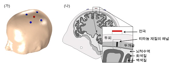
[Note] A diagram of the quasi-invasive electrical stimulation method. (A) Electrodes non-invasively attached to the scalp. (B) An illustration of a titanium channel with high conductivity (with a diameter of 1 mm)
The researchers first reconstructed the brain"s structural features using magnetic resonance imaging (MRI) and implemented brain models that attach electrodes to the scalp and implant titanium channels into the skull using 3-D brain computing.
After the neural model, which plays a central role in motor neurotransmission, is hypothetically coupled to the brain model, the influence of the brain electrical stimulation is applied through titanium channels (length: 5 mm) of various diameters (1 to 9 mm) the neuronal activation pattern according to the distance change is analyzed.
As a result, the quasi-invasive method was able to induce activation reaction of up to 11 times more neurons with about 5 times higher stimulation concentration than non-invasive methods. The titanium channel serves as an interface between the electrode and the brain, and the current applied to the electrode can be transferred to the brain without loss. (See Figure 1)
* (Supplementary explanation) In the non-invasive method of attaching electrodes to the scalp without a titanium channel, only a very small amount of current is delivered to the brain because the current applied from the electrodes spreads to the scalp, skull, and cerebrospinal fluids.

Figure 2 (a) designates the precentral gyrus of the motor cortex as the target area (ROI), (b) the electric field distribution due to noninvasive / quasi-invasive stimulation . As a result, in the case of quasi-invasive stimulation, it was confirmed that the influence of electric stimulation (red) was distributed in a relatively concentrated region compared top non-invasive stimulation. The maximum induction field strength was also higher in the quasi-invasive method (1.8 V/m) than in the non-invasive method (0.15 V/m).

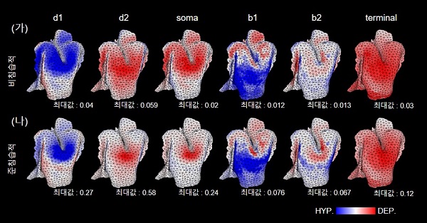
[Figure 3] Comparison of pyramidal neuron activation (brain activation area) with noninvasive / quasi-invasive stimulation. At this time, the difference (potential) (membrane potential) of the potentials varying by the external voltage is measured in the cell membrane in the dendrite (d1, d2), soma, axon (b1, b2). In the case of quasi-invasive stimulation (B), high membrane potential was observed in all regions of the neuron compared to the noninvasive stimulus (A), and observed about 10 times more activation reaction especially in the nucleus (soma). Activation by quasi-invasive stimulation (red: depolarization *, blue: hyperpolarization *) was also intensively observed near the target region.
□ Professor Sung Chan Jun said, "This study proved the utility of quasi-invasive electrical stimulation by using a computer-assisted brain stimulation model. In the future, I expect that the quasi-invasive methodology will be applied in practice to help establish a better brain stimulation strategy."

(From left) Professor Sung Chan Jun (corresponding author), Hyeon Seo graduate student (first author), Professor Hyoung-Ihl Kim (second author)
□ This research is supported by the GIST Research Institute (GRI) *, as well as by the National Research Foundation Individual Researcher Support Project, the GIST Technology Institute (GTI), and the paper was published on January 13, 2017, in Scientific Reports.
* GIST Research Institute (GRI): An organization that manages 8 research centers at GIST.